
Cell Immortalization
Cell Immortalization
Cell Immortalization Service Request FormCell Immortalization Service
Reliable and risk-free cell immortalization services to save your time and effort
Highlighted Features
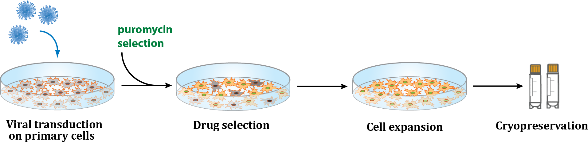
Flexible Methods
Simian virus 40 (SV40) T antigen
Human telomerase reverse transcriptase (hTERT)
Risk-Free
Invoiced only upon success
Characterization
PCR analysis of transgene expression
Timeline
2-5 months depending on immortalizing method, source cells, and species
Deliverables
5 vials (>1 x 10⁶ cells/vial) of immortalized pooled cells and/or single clone cells.
Customer Provides
Two standard vials of the desired cells
Indication of appropriate growth medium and conditions
Note: If your cells require media other than DMEM or RPMI, please provide 1–2 L of the appropriate medium along with any necessary growth factors. Additionally, supply coated 6‑well plates and T25 flasks if your cells require specially treated culture vessels.
Background

Publications
- Benischke AS et al., Sci Rep. 2017 Jul 27;7(1):6656. doi: 10.1038/s41598-017-06523-2.
Note: Restriction may apply
Have Questions?
If you don't see the product you're looking for, please don't hesitate to CONTACT US.
Our dedicated team of scientists will provide you with our best solutions for your desired results.